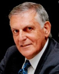
Flogen
In Honor of Nobel Laureate Prof. M Stanley Whittingham
Logo

Banner

Abstract Submission Open ! About 500 abstracts submitted from around 60 countries.


Featuring many Nobel Laureates and other Distinguished Guests

Back
    MELATONIN AND ITS METABOLITES: MAJOR CONTRIBUTORS TO THE MAINTENANCE OF CELLULAR REDOX HOMEOSTASIS - PART 2
    Russel Reiter1; Ramaswamy Sharma1; Gang Huang1;
    1UT HEALTH SAN ANTONIO, San Antonio, United States;
    PAPER: 148/Oxidative/Keynote (Oral) OS
    SCHEDULED: 11:55/Thu. 30 Nov. 2023/Dreams 1


    ABSTRACT:

    Melatonin (N-acetyl-5-methoxytryptamine) was discovered as a secretory product of the mammalian pineal gland in 1958. At the time, melatonin was thought to be exclusively synthesized and released from the pineal gland at night with the duration of the nocturnal melatonin elevation being related to the length of the daily dark period. Since the duration of the night length varies seasonally, melatonin rhythm was originally believed to regulate only circadian (sleep/wake cycle) and circannual (annual reproductive fluctuations in seasonally breeding species) rhythms, i.e., to function as both a physiological clock and as a calendar. Subsequently, melatonin was also identified in non-mammalian vertebrates, all of which have a pineal gland, and soon thereafter in invertebrates including unicells and in plants, none of which have a pineal gland [1]; thus, melatonin synthesis is obviously not exclusively of pineal origin. 

    Within the last three decades, melatonin or its synthesizing enzymes have been found in prokaryotes including true bacteria and archaebacteria (archaeans). Thus, melatonin presumably evolved in prokaryotes about 3.0-2.5 billion years ago.  During eukaryogenesis, which occurred an estimated 2.5 – 2.0 billion years ago, prokaryotes were engulfed by early eukaryotes for food and energy; the prokaryotes eventually established a symbiotic relationship with the eukaryotes that had phagocytized them, and they eventually evolved into mitochondria.  The origin of mitochondria from prokaryotes is a widely accepted theory.  The melatonin-synthesizing activity of the prokaryotes was retained in all eukaryotic mitochondria [2]. As a result, the mitochondria of many cells of present-day eukaryotes may also produce melatonin; thus, vertebrates have two sources of melatonin. The pineal gland of vertebrates discharges melatonin into the blood and cerebrospinal fluid (the releasable pool) for circadian and circannual rhythm regulation.  Additionally, all plant and animal species synthesize melatonin in their mitochondria, which is used as an antioxidant in the cell of origin (the non-releasable pool).

    Numerous publications have shown melatonin to be a multifaceted and highly efficient direct free radical scavenger and an indirect antioxidant. Melatonin quenches highly destructive reactive oxygen (ROS) and reactive nitrogen species as does many of its metabolites in what is referred to as the antioxidant cascade.  Additionally, melatonin promotes the activities of numerous antioxidant enzymes while inhibiting pro-oxidant enzyme activities. Since mitochondria are a major source of ROS generation when electrons leak from complexes I and II of the electron transport chain and chemically reduce nearby ground-state oxygen to the superoxide anion radical, melatonin synthesis in mitochondria is ideally situated to reduce free radical damage [3].  Supplemental melatonin limits elevated oxidative stress in many pathologies, such as in models of ischemia/reperfusion including stroke and heart attack, exposure of animals to heavy metals, environmental particulate matter, microplastics, hyperglycemia and atherosclerosis, excessive alcohol, conventional medications including chemotherapies, illegal drugs, ionizing radiation, neurodegenerative diseases, and those caused by dysfunctional mitochondria. These studies have been extended to humans where melatonin has similar protective actions. Some of the functions of melatonin involve transmembrane receptors which are widely distributed while other actions are receptor-independent [4].                                            


    References:
    [1] D. Zhou, Y, Yu, J. Zhang, R. Zhang, Z, Mou, X. Zhang, Z. Li, X. Feng, S. Chen, R.J. Reiter. Front. Endocrinol. 10 2019) 19249.
    [2] D.X. Tan, L.C. Manchester, Z. Liu, S.A. Rosales-Corral, D. Acuna-Castroviejo, R.J. Reiter. J. Pineal Res., 54 (2013) 127-138.
    [3] R.J. Reiter, S.A. Rosales-Corral, D.X. Tan, M.J. Jou, A. Galano, B. Xu. Cell. Mol. Life Sci. 74 (2017) 3863-3881.
    [4] R.J. Reiter, Q. Ma, R. Sharma. Physiology (Bethesda) 35 (2020) 86-95.