In Honor of Nobel Laureate Prof. M Stanley Whittingham


 |
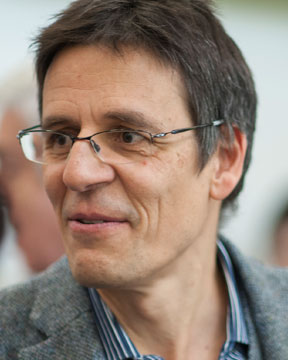
Joachim FrankColumbia UniversityCapturing Motion Of Biomolecules By Single-particle Cryo-em Summit Plenary Back to Plenary Lectures » |
Abstract:Cryogenic electron microscopy (cryo-EM) has transformed the way we can study biological molecules as it enables us to determine structure of molecules freely suspended in solution (as “single particles”), and to gauge the way they change their shape as they interact with one another in the cell. Freezing, by plunging the sample into a cryogen at liquid nitrogen temperature, is necessary to trap the molecules in a fixed position during imaging and to reduce the damage inflicted by the electrons in the process. Following the introduction of novel cameras for detecting single electrons, near-atomic resolution has been achieved for many molecules and molecular machines of biomedical relevance, including ion channels and receptors. Most recently cryo-EM has contributed in major ways to the development of vaccines and cures for COVID-19. The standard protocol of cryo-EM does not allow visualization of reactions since the time for sample deposition on the grid and blotting is in the range of several seconds. To study the process of a reaction, as it goes through one or more short-lived intermediates, we have developed a method of time-resolved cryo-EM. Here two reactants are mixed in a microfluidic channel, allowed to react for a defined time (between 10 and 1000 milliseconds), and the reaction product sprayed onto the EM grid as the latter is being plunged into the cryogen. With this approach we have been able to visualize short-lived states of the ribosome in different stages of its work cycle making proteins. Applications in the study of many other molecular machines are possible. |
|